 Safekipedia
Golgi apparatus
Adapted from Wikipedia · Adventurer experience
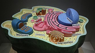
The Golgi apparatus, also called the Golgi complex, Golgi body, or simply the Golgi, is a tiny part inside most eukaryotic cells. It lives in the cytoplasm. Its job is to pack proteins into tiny bags called membrane-bound vesicles. These bags then go to where they are needed in the cell.
The Golgi apparatus helps prepare proteins for secretion. It uses special tools called glycosylation enzymes to add sugar pieces to proteins.
This structure was first seen in 1898 by an Italian scientist named Camillo Golgi. It was later named after him in the 1910s.
Discovery
The Golgi apparatus was one of the first tiny parts inside cells to be found. An Italian doctor named Camillo Golgi discovered it in 1898. He was looking at the nervous system with his microscope. At first, some people thought it might just be a trick of the microscope. But with better microscopes, scientists learned that the Golgi apparatus really exists. It was called different names before it became known as the Golgi apparatus.
Subcellular localization
The Golgi apparatus is found in different places inside cells, depending on the cell type. In animals, there is usually one Golgi apparatus near the center of the cell, close to an area called the centrosome. In yeast cells, many smaller Golgi apparatuses are spread out. In plant cells, the Golgi stacks are not in one spot and do not form ribbons. All of them are next to areas where the endoplasmic reticulum sends out material.
Structure
In most cells, the Golgi apparatus is made of flattened, disk-like parts called cisternae. These come from small bubbles that break off from another part of the cell called the endoplasmic reticulum.
The Golgi is divided into different parts, called the cis Golgi network and the trans Golgi network. The cis part is where the process starts, and the trans part is where it ends, packing proteins into small bags called vesicles. These vesicles then carry proteins to other parts of the cell or to the outside.
The size and number of Golgi structures can vary depending on the cell. Cells that make and release a lot of substances have larger and more noticeable Golgi structures. Each stack of cisternae has an entry side and an exit side, and they contain special enzymes that change the proteins, helping decide where each protein goes next.
Function
The Golgi apparatus works like a busy post office inside cells. It takes proteins made in a part of the cell called the endoplasmic reticulum and puts them into small packages called vesicles. These vesicles go to the Golgi apparatus, where the proteins are changed and ready for their next job. Some proteins leave the cell, while others stay to help the cell.
The Golgi apparatus also helps move fats called lipids and makes special parts of the cell called lysosomes. It changes proteins by adding sugar molecules and other groups. These changes help the proteins know where to go next in or around the cell. The Golgi also helps build important molecules called proteoglycans that are found outside the cell.
Vesicular transport
Tiny bags of material from the rough endoplasmic reticulum move to the cis face of the Golgi apparatus. They join with the Golgi's membrane and let their contents inside.
Inside, the molecules are changed and then sorted to go to their next places in the cell.
Proteins that need to leave the endoplasmic reticulum or the Golgi move through the Golgi to the trans face. There, they reach a special area called the trans-Golgi network (TGN). This is where proteins are sorted and sent to where they need to go, placed into different types of bags based on the signals they carry.
| Types | Description | Example |
|---|---|---|
| Exocytotic vesicles (constitutive) | Vesicle contains proteins destined for extracellular release. After packaging, the vesicles bud off and immediately move towards the plasma membrane, where they fuse and release the contents into the extracellular space in a process known as constitutive secretion. | Antibody release by activated plasma B cells |
| Secretory vesicles (regulated) | Vesicles contain proteins destined for extracellular release. After packaging, the vesicles bud off and are stored in the cell until a signal is given for their release. When the appropriate signal is received they move toward the membrane and fuse to release their contents. This process is known as regulated secretion. | Neurotransmitter release from neurons |
| Lysosomal vesicles | Vesicles contain proteins and ribosomes destined for the lysosome, a degradative organelle containing many acid hydrolases, or to lysosome-like storage organelles. These proteins include both digestive enzymes and membrane proteins. The vesicle first fuses with the late endosome, and the contents are then transferred to the lysosome via unknown mechanisms. | Digestive proteases destined for the lysosome |
Current models of vesicular transport and trafficking
Scientists are still learning how proteins move through the Golgi apparatus. There are a few ideas about how this happens.
One idea is that the Golgi has stable sections. Each section has special enzymes that change proteins. Proteins move between sections in tiny bags called vesicles. Another idea is that these sections change over time, and proteins move as the sections mature. A newer idea is that the Golgi has stable sections controlled by special proteins called Rab GTPases. Each idea has good points and bad points, and scientists are still figuring out how proteins move through the Golgi.
Brefeldin A
Brefeldin A (BFA) is a substance made by fungi. Scientists use it to study the Golgi apparatus. When added to cells, BFA stops some proteins from working. This makes the Golgi apparatus break apart and its proteins move to other parts of the cell. This helps researchers learn more about how cells send out materials.
Images
Related articles


This article is a child-friendly adaptation of the Wikipedia article on Golgi apparatus, available under CC BY-SA 4.0.
Images from Wikimedia Commons. Tap any image to view credits and license.